
Methods for Weld Penetration, Structure Observation, and Defect Analysis
焊接是一种用于加入金属的技术,在各种领域中,包括植物设备和汽车领域。焊接穿透,基材和珠子可能患有由长期使用的压力或腐蚀引起的缺陷。因此,重要的是观察和分析焊接部件的表面和横截面。本节介绍该方法,以及使用我们最新的4K数字显微镜的观察和分析示例,大大改变了传统显微镜的观察和分析的概念。
- 焊接渗透分析的重要性
- 观察和分析焊接渗透(金属结构)的方法
- Latest Examples of Weld Penetration Observation and Analysis
- New Common Practices of Weld Penetration Observation and Analysis
焊接渗透分析的重要性
其他金属连接技术,焊接是一个rational, airtight fabrication process used in a wide range of applications. Infrastructure, architecture, automobiles, electrical appliances, and electronic devices all employ a variety of welding methods. These different methods, such as variations of arc welding, laser beam welding, and resistance spot welding, are utilized according to purpose and base material.
特别是,工厂的工艺设备旨在用于几十年。这一要求要求焊接质量最高。有许多类型的植物,包括热电厂;新材料由石油制成的石油化工厂;制造一般化学品,药物和食物的植物;用于净化污水的水处理厂;和工业废物处理厂进行再循环过程。所有这些植物都是巨大的设施,需要具有不同性质和不同状态的液体和气体。当在焊接时检测到老化或缺陷时,设备的观察和分析对于识别问题和原因是至关重要的。这通常不是一项简单的任务。
观察和分析焊接渗透(金属结构)的方法
In the case of large-scale facilities such as plants, micro analysis cannot be performed on the welded structure. However, defects such as poor fusion, slag inclusion, or cracks (stress corrosion cracks and fatigue cracks) in the welded part still require investigation. Therefore, the majority of microscopic observation and analysis of these issues employ the following two types of sample creation methods:
- Microscopic observation of sample filled with resin (embedment)
- The welded part with the defect is cut out as a sample and is filled (embedded) with resin for microscopic observation. Since the sample is cut out, it is possible to observe the cross-section of the weld penetration.
- 通过贮槽进行显微镜观察
- “sump”代表Suzuki的通用微型印刷。如果不能为显微镜观察切出样品,则样品的表面被转移到纤维素板上,并使用现场显微镜观察。据说用于电子显微镜的复制样品源自贮槽。
这些观察方法涉及模拟样品的观察方法不仅用于植物,而且还与汽车,航空有关的设备,铁路车辆和船舶一起使用。总之,可以找到任何地方的焊接故障,缺陷会导致主要事故。在每种情况下,使用表面和横截面观察观察和分析焊接部分的金属结构;以及其他技术。
Latest Examples of Weld Penetration Observation and Analysis
由于近年来技术进步,数字显微镜已经解决了传统显微镜的问题,显着改变了倍率清晰度,并提高了样品的可加工性,用188金宝搏下载app于观察和分析焊接渗透。
Keyence的VHX系列超高清4K数字显微镜采用最新的高分辨率镜头,4K CMOS,照明选项和图像处理技术,从而能够通过与传统显微镜进行比较,通过清晰的图像和理性分析进行观察。
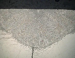
Cross-section observation and defect analysis of weld penetration structure
通过传统的显微镜,在观察中只有一部分焊缝穿透的金属结构是焦点,这就是该程序需要很长时间的原因。熟练程度也需要实现准确的观察和分析。
The VHX Series 4K Digital Microscope allows for quick and easy capture of fully focused images by using depth composition. These fully-focused images contribute to clear observation of the condition of the metal structure of the weld penetration and of fine cracks in the base material, regardless of the user's skill level, thereby helping to prevent problems from being overlooked.


Weld penetration structure observation using SUMP samples
With conventional microscopes, samples created by SUMP may only partially come into focus due to the need for high-magnification observation. Additionally, replicas have low contrast, which can make observation impossible.
使用清晰的高分辨率4K图像,VHX系列可以观察和分析具有不均匀表面和低对比度的块样本。

Achieving on-site micro analysis with handheld observation
Conventionally, large test targets could not be observed under a microscope on site—the only ways to observe and analyze such targets were to cut out a sample and fill it with resin (embedment) or, if a sample could not be cut out, use SUMP to transfer or create a replica. The accuracy and workability of the investigation were also affected by the quality of the created samples.
The VHX Series 4K Digital Microscope can perform observation and analysis in handheld form, providing clear magnified images produced by the deep depth of field of the dedicated lens. This observation and analysis can eliminate the time, work, and hassle of creating samples as well as enable non-destructive observation and analysis of the actual target on the spot.

A single unit supports various 2D and 3D measurements
VHX系列4K数字显微镜不仅可以执行明显的放大率和观察,而且还可以进行传统显微镜的测量和量化。
2D和3D测量,粗糙度测量,技术清洁度检测,晶粒尺寸测量和检查金属结构和产品的其他有用功能,可以使用简易鼠标操作从单个机器中容易地执行。

New Common Practices of Weld Penetration Observation and Analysis
焊缝和金属结构的观察和分析非常重要,因为这些操作与各种金属产品和设备的安全密切相关。识别和分析问题需要准确性和速度。
The VHX Series is equipped with many other advanced functions that were not conventionally available. Automatic zoom from 20x to 6000x, multi-lighting functionality, and 2D and 3D measurements contribute to advanced observation and analysis that is not possible with conventional microscopes.
For additional product info or inquiries, click the buttons below.
